View Cores Alphabetically
FIND A CORE FACILITY TO HELP WITH YOUR RESEARCH
Search below to explore services, equipment, locations, service categories and more
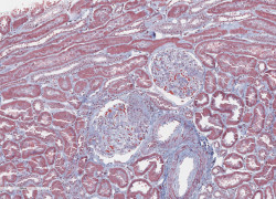
Digital Pathology Core
Peter D. Ouillette
734-764-4003
[email protected]
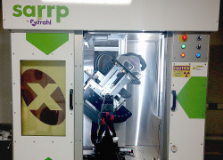
Experimental Irradiation Core
Meredith Morgan, PhD
734-647-5928
[email protected]
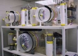
Germ-Free Mouse Facility
Germ-Free Mouse Facility
734-615-9420
[email protected]
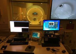
Human Research 3 Tesla Magnetic Resonance Imaging (Research 3T)
Thomas L. Chenevert, PhD
734-936-8866
[email protected]
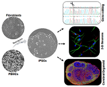
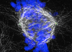
Human Stem Cell and Gene Editing Core (HSCGE)
Rouknuddin Ali
[email protected]

Institute for Healthcare and Policy Innovation (IHPI)
Patrick Brady, MHA
734-763-4335
[email protected]
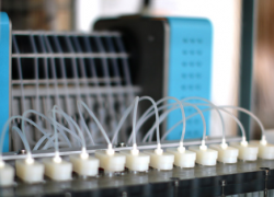
Metabolic, Physiological and Behavioral Phenotyping Core (MMPC-Live)
Nathan Qi, MD/PhD
(734) 764-7043
[email protected]
Michigan Diabetes Research Center (MDRC) Clinical Core
William Herman, MD, MPH
734-936-8279
[email protected]

Michigan Diabetes Research Center (MDRC) Islet Core
Corentin Cras-Meneur, PhD
734-232-8165
[email protected]
Michigan Diabetes Research Center (MDRC) Microscopy & Image Analysis Core (MIAC)
David A. Antonetti, PhD
734-232-8230
[email protected]
Michigan Diabetes Research Center (MDRC) Molecular Genetics Core (MGC)
David P. Olson, MD, PhD
734-232-8205
[email protected]
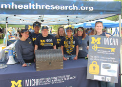
Michigan Institute for Clinical & Health Research (MICHR)
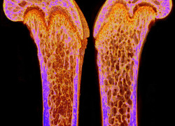
Michigan Integrative Musculoskeletal Health Core Center (MiMHC)
Karl Jepsen, PhD
[email protected]
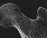
Micro & Nano Computed Tomography Advanced Imaging Core
Andrea Clark
734-615-6956
[email protected]
Microbiome Core
Tom Schmidt, Ph.D
[email protected]
Miller Lab
Melissa Han
734-936-2164
[email protected]

Mixed Methods Program
Satoko Motohara
(734) 936-5672
[email protected]

Morphomic Analysis Group (MAG)
June Sullivan
734-764-7841
[email protected]
Neuropsychology Program
Carol Persad, PhD
734-763-9259
[email protected]
Total Cores: 51
Total Cores: 51