Human Research 3 Tesla Magnetic Resonance Imaging (Research 3T)
provides anatomic and advanced MR imaging services for IRB-approved, sponsor-funded studies and clinical trials.
Contacts
Thomas L. Chenevert, PhD
734-936-8866
[email protected]
Location
University of Michigan Hospital (Rm B2B405)
1500 East Medical Center Drive, Ann Arbor, MI
Medical School
Who We Serve
University of Michigan Researchers and External Researchers
Core Summary
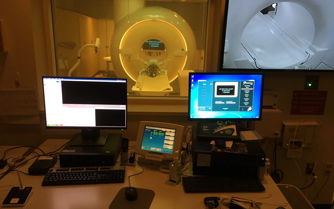
The Department of Radiology Research 3 Tesla MRI facility located on the University Hospital B2-Level provides high quality anatomic and advanced MR imaging services for IRB-approved, sponsor-funded studies and clinical trials. The research scanner, a 3T Philips Ingenia operating at software version 5.6x, offers a 70cm diameter wide bore for subject comfort. The system is fully digital with on-coil conversion to optical, patient-specific dual-channel transmit B1 shimming and second-order B0 shimming. The system contains a full complement of coils including: 32channel head; 15ch head; 20ch head & neck; 52ch total neuro; two 16ch anterior torso/cardiac for whole-body studies; 16ch breast and 7ch for breast-biopsy; and all MSK coils (knee, shoulder, wrist, small extremity, foot/ankle, and three flex coil pairs). Each gradient axis provides 40mT/m amplitude @200 T/m/s slew; or 78mT/m amplitude @100 T/m/s slew and 100% duty cycle. The research scanner is operated by two RT(R) certified research-dedicated MRI Technologists for safety screening, medical-grade handling and scanning of research subjects. The facility is also supported by research faculty skilled in MRI acquisition, image/data processing and customization of scan protocols to meet study requirements and can assist with HIPPA-compliant image/data transfer to UM investigators or external research core labs.
Services
- Diffusion weighted and diffusion tensor imaging
- Expert consultation on image acquisition/analysis procedures
- High spatial-/temporal anatomic imaging
- MR angiography and venography
- MR elastography
- Quantitative imaging: fat-content and relaxometry
- Single- and multi-voxel proton spectroscopy
- Tissue perfusion/vascular permeability via DSC/DCE/ASL
- Tissue/organ kinetic studies
Equipment
- Expression’s Patient Monitor
- Medrad MRX Power Injector
- Philips 3 Tesla MRI Model Ingenia SW R5.4
- SensaVue II Entertainment and fMRI system