Histology, Pathology and Tissue
FIND A CORE FACILITY TO HELP WITH YOUR RESEARCH
Search below to explore services, equipment, locations, and more
Central Biorepository (CBR)
Provides access to and storage of high-quality, highly annotated human biospecimens.
Custom kit production and biospecimen chain of custody from clinic-CBR-end user.
Victoria Blanc, PhD
734-763-6423
[email protected]
Dental School Histology Core Facility
specializes in sectioning of hard (demineralized) tissues and offers basic histology and staining.
Theresa Cody
(734)764-1543
[email protected]
Digital Pathology Core
generates digital representations of glass microscope slide tissue (whole-slide images) in “SVS” format.
Peter D. Ouillette
734-764-4003
[email protected]
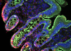
Michigan Diabetes Research Center (MDRC) Microscopy & Image Analysis Core (MIAC)
provides protein/RNA imaging and analysis for diabetes-related research
David A. Antonetti, PhD
734-232-8230
[email protected]
Michigan Institute for Clinical & Health Research (MICHR)
educates, funds, connects & supports clinical and translational research teams.
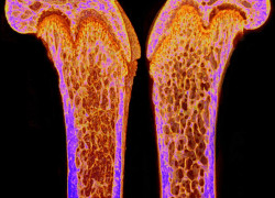
Michigan Integrative Musculoskeletal Health Core Center (MiMHC)
Our 3 Cores provide analyses aimed at understanding musculoskeletal health using paraffin and plastic (hard tissue) histology and training, micro/nanoCT imaging, Raman spectrometry, whole animal/tissue level testing, Omics and machine learning support.
Karl Jepsen, PhD
[email protected]
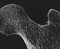
Micro & Nano Computed Tomography Advanced Imaging Core
provides 3-D imaging and quantitative analysis of structures/materials, including metals, silica-based chips and plastics.
Andrea Clark
734-615-6956
[email protected]
Platelet Pharmacology and Physiology Core
facilitates blood collection, pharmacological equipment usage and training services.
Amanda Prieur
734-763-8824
[email protected]
Rogel Cancer Center Tissue and Molecular Pathology (TMP) Core
provides tissue procurement, histology, and molecular pathology services.
Thomas Giordano, MD, PhD
[email protected]
ULAM Pathology Core (formerly IVAC)
is a research pathology core run by ULAM providing histology, bloodwork, pathology, and technical study support on a fee-for-service basis.
Ingrid L. Bergin, VMD, MS, DACLAM, DACVP
(734) 936-3395
[email protected]
Total Cores: 10
Total Cores: 10