Rogel Cancer Center Tissue and Molecular Pathology (TMP) Core
provides tissue procurement, histology, and molecular pathology services.
Contacts
*General Questions
734-647-3261
Path-TMPSR-Histology@med.umich.edu
Dafydd Thomas, MD, PhD
thomasda@med.umich.edu
Location
MPRL and Tissue Procurement: Medical Sciences Research Building I, Room 4500
Histology Core: Medical Sciences Research Building I, Room 4504
1150 W. Medical Center Drive, Ann Arbor, MI
Medical School
Who We Serve
University of Michigan Researchers
Core Summary
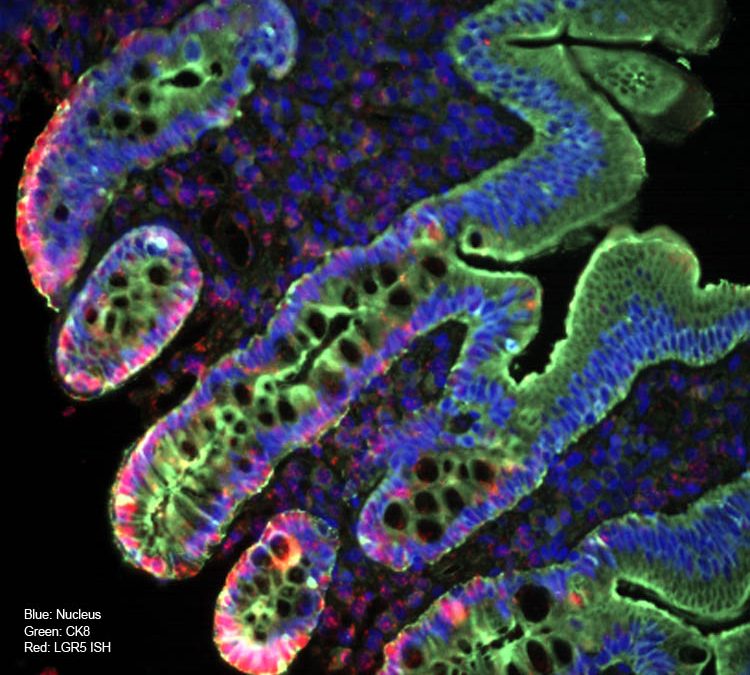
The Rogel Cancer Center Tissue and Molecular Pathology (TMP) Shared Resource (SR) Consists of three distinct, yet complimentary services relating to the procurement and evaluation of tissue for research: Tissue Procurement, Histology and Molecular Pathology. The Tissue Procurement service provides pathology-sanctioned procurement of surgically resected neoplastic and non-neoplastic tissue (fresh and frozen). The Histology core provides sample processing, embedding, sectioning, routine H&E, special staining, immunohistochemistry (IHC) and frozen sectioning, as a fee-for-service core. The Molecular Pathology Research Laboratory (MPRL) performs IHC, in-situ hybridization, multiplex immunofluorescence, FISH staining and tissue microarray construction, as a fee-for-service core. We can perform staining on frozen tissue or FFPE, using DAKO or Ventana autostainers. In addition to these services, TMP Shared Resource provides multispectral and brightfield whole slide imaging using a Polaris scanner, to facilitate digital analysis of immunofluorescence and chromogenic immunohistochemistry projects.
Services
- Frozen sectioning
- Hematoxylin and eosin staining
- Immunofluorescence (including multiplex)
- Immunohistochemistry
- Molecular pathology services
- Special stains
- Tissue banking
- Tissue microarray construction
- Tissue processing/embedding/sectioning
- Vectra Polaris whole-slide multispectral and brightfield scanning (whole slide imaging)
Equipment
- Autostainer XL
Leica, Core Use Only - CM1860 Cryostat
Leica, Core Use Only - CY5030 Coverslipper
Leica, Core Use Only - Discovery Ultra
Ventana, Core Use Only - Histocore Arcadia C
Leica, Core Use Only - Histocore Arcadia H
Leica, Core Use Only - Histocore BioCut
Leica, Core Use Only - Stainer Autostainer Link48
Dako/Agilent, Core Use Only - Whole slide scanner Vectra Polaris
Akoya, Core Use Only


