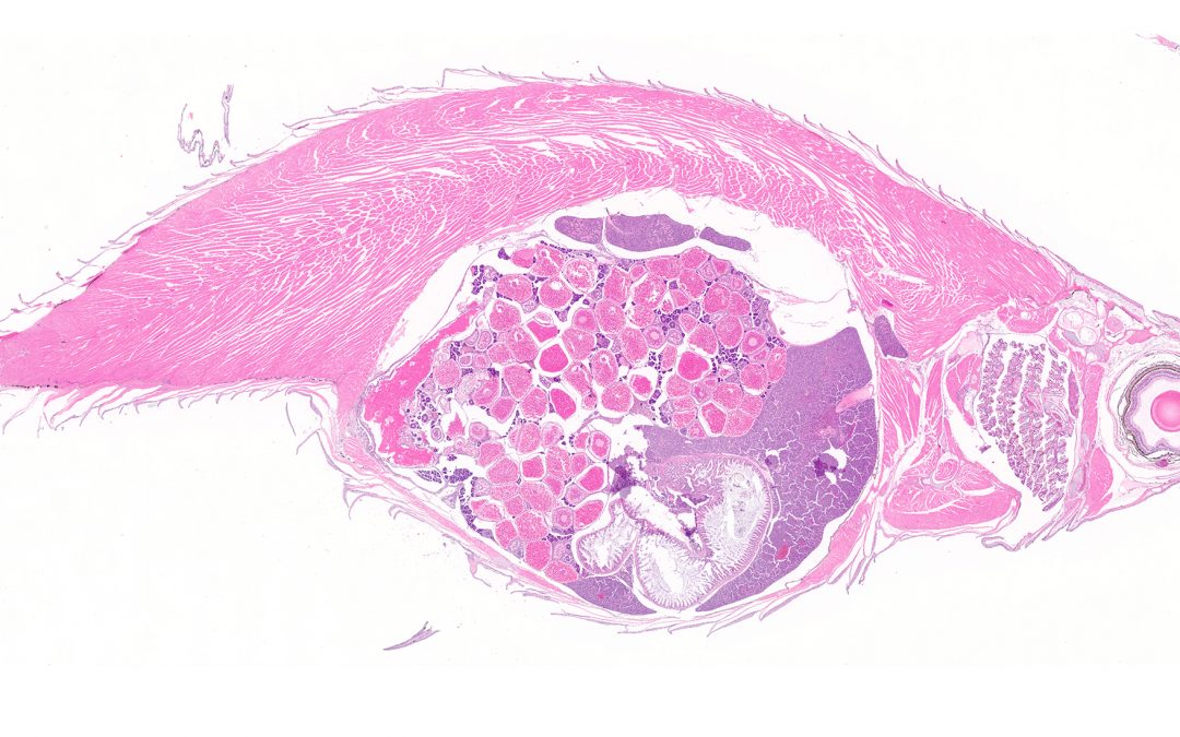
ULAM Pathology Core (formerly IVAC)
is a research pathology core run by ULAM providing histology, bloodwork, pathology, and technical study support on a fee-for-service basis.
Contacts
Ingrid L. Bergin, VMD, MS, DACLAM, DACVP
(734) 936-3395
ULAM-PathologyCore@umich.edu
Location
North Campus Research Complex
Building 36-G155/157
2800 Plymouth Road
Ann Arbor, MI 48109
(734) 936-3395
SATELLITE: 3527 ARF
1150 W. Medical Center Drive
Ann Arbor, MI 48109
(734) 936-3803
Medical School
Who We Serve
University of Michigan Researchers and External Researchers
Core Summary
- Necropsy/tissue collection
- Histology/immunohistochemistry
- Animal hematology/clinical chemistry
- Pathology interpretation and quantitative image analysis
- Digital slide scanning (brightfield)
- Project consultation
Services are provided on a fee-for-service basis, and estimates/pre-project consultations are available and encouraged. ULAM Pathology Core submissions can be made by appointment through MiCORES
ULAM Pathology Core’s main lab is located at NCRC, but a convenient satellite location on the Medical Campus with courier service between the two labs is also available.
Additionally, the Technical Services Team can provide support for in-vivo studies via assistance with dosing, monitoring, blood/tissue collection, ear tagging/other animal identification, and training on specific techniques. Submit Tech service requests through our website.
Services
- Animal diagnostic laboratory (ADL) testing
- Blood collection
- Clinical chemistry
- Digital image analysis
- Digital slide scanning
- Dosing
- Hematology
- Histology
- Necropsy
- Pathology evaluation and reporting
- Quantitative morphometry
Equipment
- Aiforia platform
Aiforia, Core Use Only - Aperio digital whole slide scanner Aperio AT2
Leica, Core Use Only - Brightfield & epifluorescent microscopes Various
Olympus, Core Use Only - Cryostat - microtome CM3050 S
Leica, Core Use Only - Down-draft necropsy tables and backdraft grossing stations for animal necropsy and tissue trimming, BSL1 and BSL2 space Various
Mopec, Core Use Only - Heska hematology analyzer Element HT5
Heska, Core Use Only - Intellipath immunohistochemistry autostainer Intellipath
Biocare, Core Use Only - Leica histostainer ST5010 XL
Leica, Core Use Only - Liasys clinical chemistry instrument Liasys 330
AMS Diagnostics, Core Use Only - Paraffin sectioning microtomes (multiple) RM2125 and others
Leica, Core Use Only - Tissue embedding station
- Tissue processors Tissue-Tek VIP 6
Sakura Finetek, Core Use Only - Type II Biosafety cabinet SCPlus
Allentown, Core Use Only

