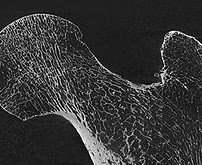
Micro & Nano Computed Tomography Advanced Imaging Core
provides 3-D imaging and quantitative analysis of structures/materials, including metals, silica-based chips and plastics.
Contacts
Andrea Clark
734-615-6956
[email protected]
Location
BSRB (Rm 2231)
109 Zina Pitcher Pl, Ann Arbor, MI
Medical School
Who We Serve
University of Michigan Researchers and External Researchers
Core Summary
The nano- and micro-Computed Tomography imaging core provides services for acquiring and quantitative analysis of three-dimensional images of biological and engineered materials, including metals, silica-based chips, scaffolds, and plastics. The core is capable of imaging mineralized tissues (e.g., bone, teeth); as well as soft tissues (e.g., tendons, ligaments, cartilage, blood vessels, muscle) with the aid of radiographic contrast agents. Images can be acquired for both ex vivo and in vivo (microCT only) studies. This core is managed and operated by members of the Orthopaedic Research Laboratories.
Services
- CT Image analysis
- Ex vivo CT Scanning
Micro CT, Nano CT,
Equipment
- In vivo/Bruker Skyscan 1176 high resolution micro-CT imaging system 1176
Bruker, Equipment Available For Use - Phoenix x-ray | Nanotom m
GE Sensing & Inspection Technologies, Core Use Only

